hiatal hernia pediatric radiology
The 36 returned revealed that the larger departments see an average of 32 hiatal hernias per year or about 062 per cent of their upper gastrointestinal studies. What are the treatment options for a hiatal hernia.

Pediatric Hiatal Hernia Radiology Case Radiopaedia Org
Repair can usually be performed laparoscopically.

. Image-guided intervention can be used to aid the surgeon in the. The obstruction which is complete or nearly so almost always appears to be the direct result of an acute volvulus of the stomach 5 13 16. Esophageal surgery is a common and integral component in the management of hiatal hernias esophageal carcinoma and esophageal perforation.
An upper GI contrast study allows definite diagnosis. See part of the stomach sliding through the esophageal hiatus into the chest DDX. A hiatal hernia happens when part of your stomach pushes up into an opening hiatus in your diaphragm.
Diaphragmatic hernias alternative plural. There are two main types of congenital diaphragmatic hernia CDHs which are uncommon yet distinct entities that usually occur on the left side 80 of the diaphragm 12. Hiatal hernia radiology discussion including radiology cases.
Congenital diaphragmatic hernia CDH recurrence secondary hiatal hernia radiologic screening longitudinal follow-up risk factors for recurrence cone-shaped patch Introduction Congenital diaphragmatic hernia CDH is a rare malformation and surgical repair is still an intervention with a remarkable complication rate. You may not need surgery. When the muscle of the diaphragm weakens the opening becomes large.
However it is commonly defined as any herniation of elements in the abdominal cavity through the esophageal hiatus of the diaphragm 2 3. A hiatal hernia occurs when a portion of the stomach moves up into the chest cavity. Paraesophageal hernia Large hernias can cause symptoms and with progressive hiatal widening increasing protrusion and rotation of the stomach can lead to gastric volvulus that can be complicated by hemorrhage obstruction strangulation perforation.
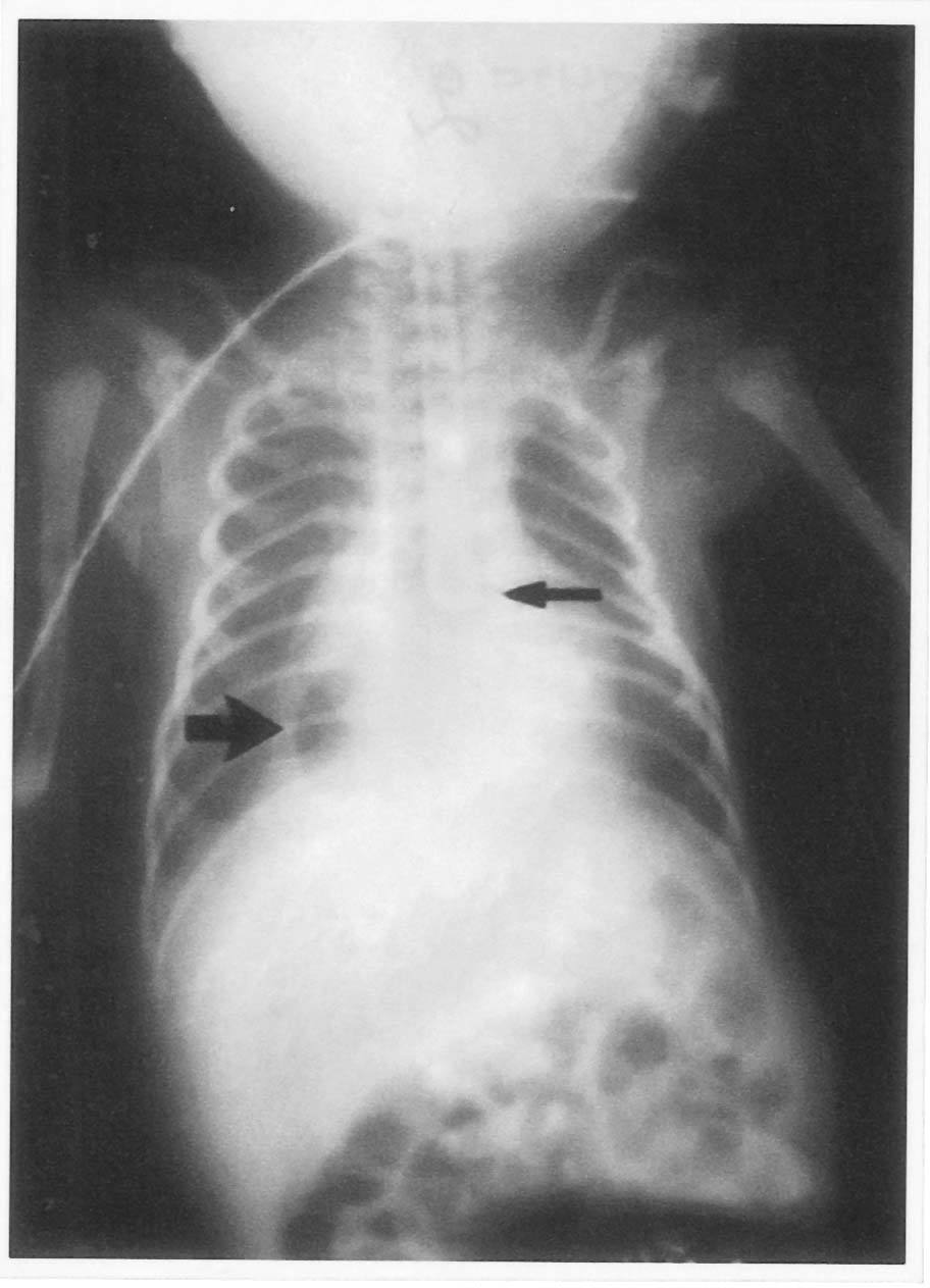
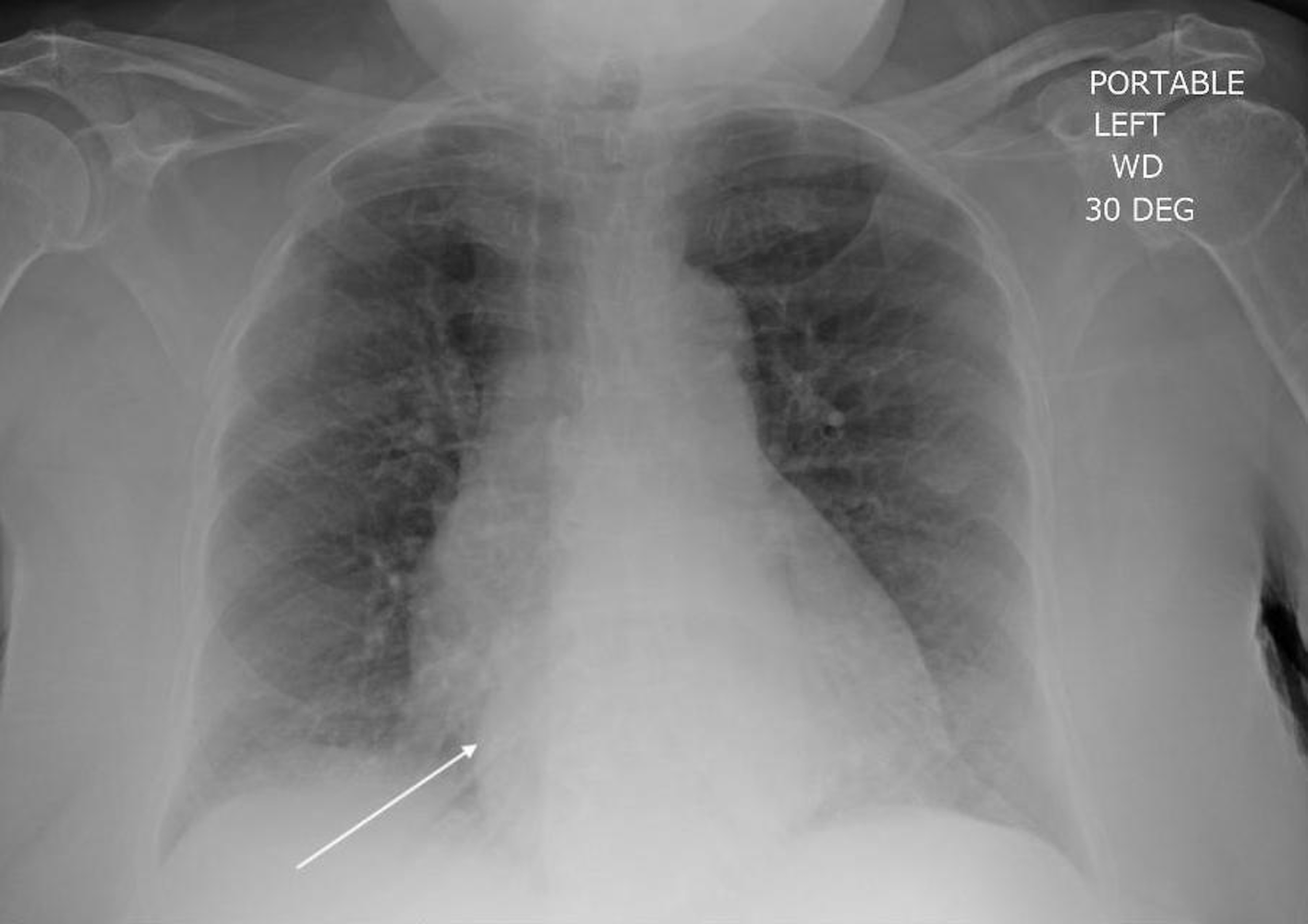
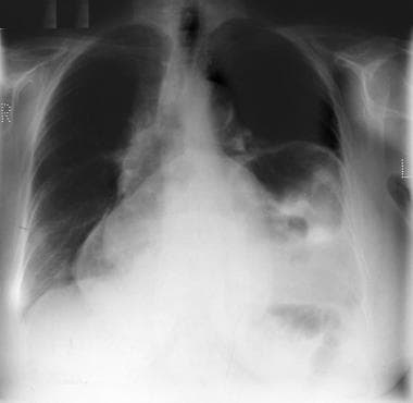
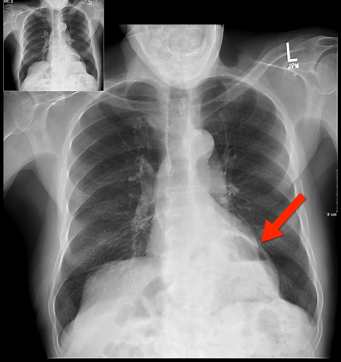
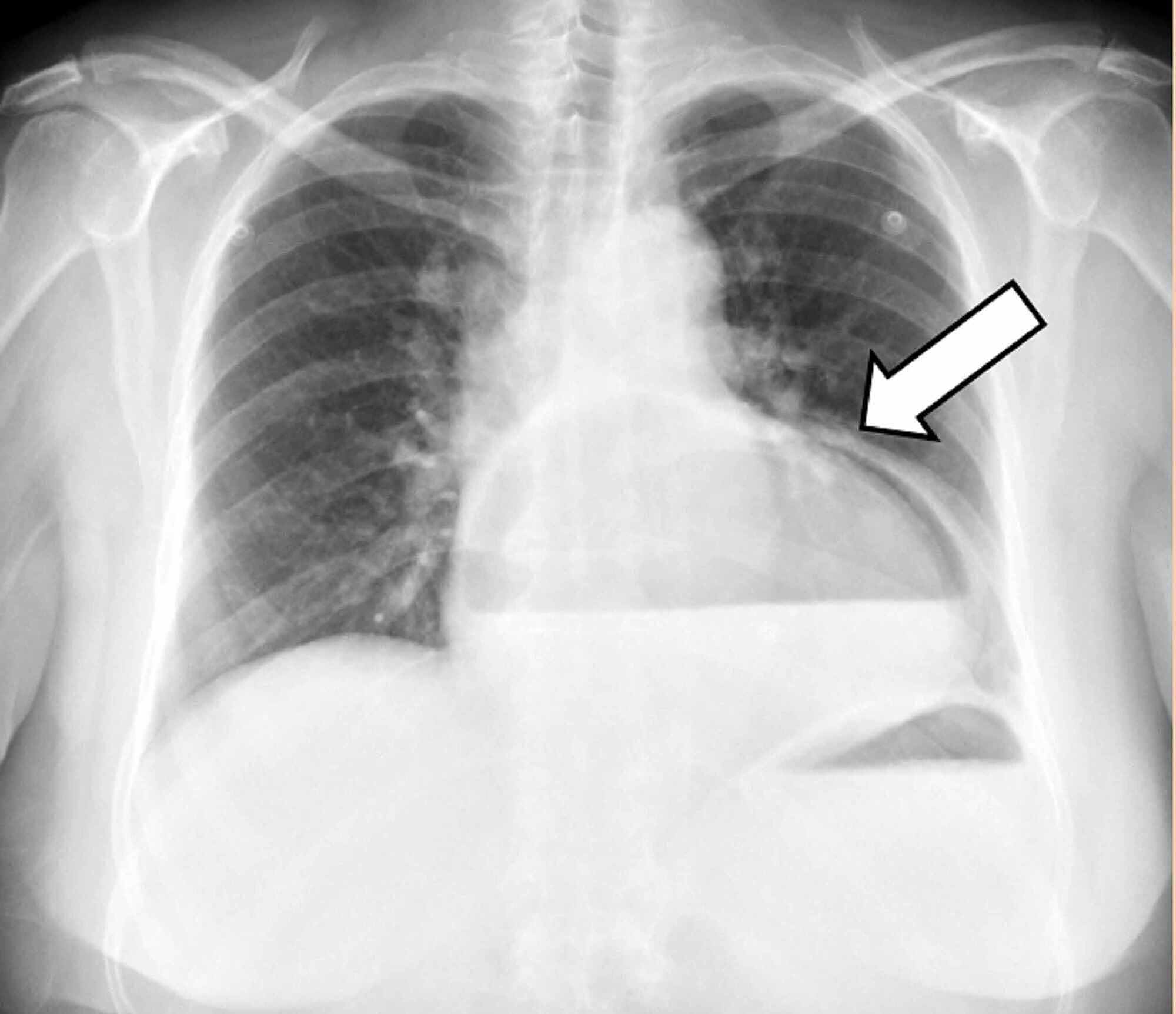
Normally the opening in the diaphragm only contains the lower part of the esophagus. At radiography hiatal hernias may be seen as a solid or air-filled retrocardiac mass. The diaphragm is the muscle that separates the chest from the abdomen.
This is often a laparoscopy but in some cases you may need open traditional surgery. These studies are usually not being done to specifically diagnose a hiatal hernia but rather to exclude other diagnoses or as part of other workup. Clinical and radiological aspects along with postoperative complications are reviewed.
Right Esophagram in a patient with type I sliding HH shows the lower esophageal sphincter or phrenic ampulla marked by the A ring proximally and the B ring distally. Persistent dysphagia occurred in 13 647 over the first year which resolved after a single balloon dilation in 67 46. Asymptomatic hiatal hernias do.
A questionaire regarding the incidence of hiatal hernia in infacts and children was sent to the 50 largest departments of pediatric radiology in the United States and Canada. A natural hole in the diaphragm allows the esophagus to pass from the chest to the abdomen. The 24-hour pH monitoring was performed as follows.
Department of Radiological Sciences. Demographics and etiology Congenital. Hiatal hernia HH in children is a well-recognized finding commonly diagnosed by both radiologists and gastroenterologists 1.
In hiatal hernia the obstruction is generally a complication of a large sliding hernia with an esophagus of normal overall length that enters the stomach above the diaphragm. Type III is the 2nd most common type but it is rare compared to type I sliding HH. 0 public playlist include this case.
Hiatal hernia is rare and causes gastrointestinal symptoms more frequently. This case was submitted with supervision and input from. The pH was measured and recorded using a pH recorder model Digitrapper MK 3 Synectics Medical AB Stockholm Sweden.
But if your case is serious you will need a hiatal hernia repair. The prevalence of HH varies widely due to inconsistency in the definition. The diaphragm separates the chest cavity from the abdominal cavity.
Hiatal hernia occurs when part of the stomach and sometimes other organs of the abdominal cavity slide upward through an opening in the diaphragm into the chest cavity. The following tests for demonstrating hiatal hernia or gastroesophageal reflux were employed on each patient. A giant mixed hiatal hernia was noted during surgery.
Following the ingestion of 8 ounces of barium the patient was placed in the supine Trendelenburg position at least 15 and the esophagogastric junction was observed. David Geffen School of Medicine at UCLA. Herniae are defined as either congenital or acquired defects in the diaphragm.
Hiatal hernia andor gastroesophageal reflux are studied in 182 pediatric patients after surgical correction of esophageal atresia. Special stress is given to late stenosis that does not cure with conventional treatment and are originated by undiagnosed reflux. Hiatal hernias are often incidentally diagnosed on radiographic imaging upper endoscopy or esophageal manometry.
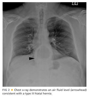
Type I is a sliding HH and types II-IV are paraesophageal hernias. Gastroesophageal reflux Cases of Hiatal Hernia CXR AP shows an air fluid level at the level of the diaphragm. When the ligaments holding the esophagus in position becomes loose the hole becomes too large.
While in the supine position the patient was. In the pediatric population Bochdaleks hernia and Morgagnis hernia are the most frequent congenital diaphragmatic hernias. The cause of hiatal hernias is unknown but children with this condition are usually born with it.
Because children with a hiatal hernia usually have concomitant gastroesophageal reflux fundoplication is usually performed at the same time. The diaphragm is a muscle between your stomach and your chest. Hiatal hernia was diagnosed as an upward displacement of the lower esophageal sphincter or identification of more than three gastric mucosal folds above the diaphragm.
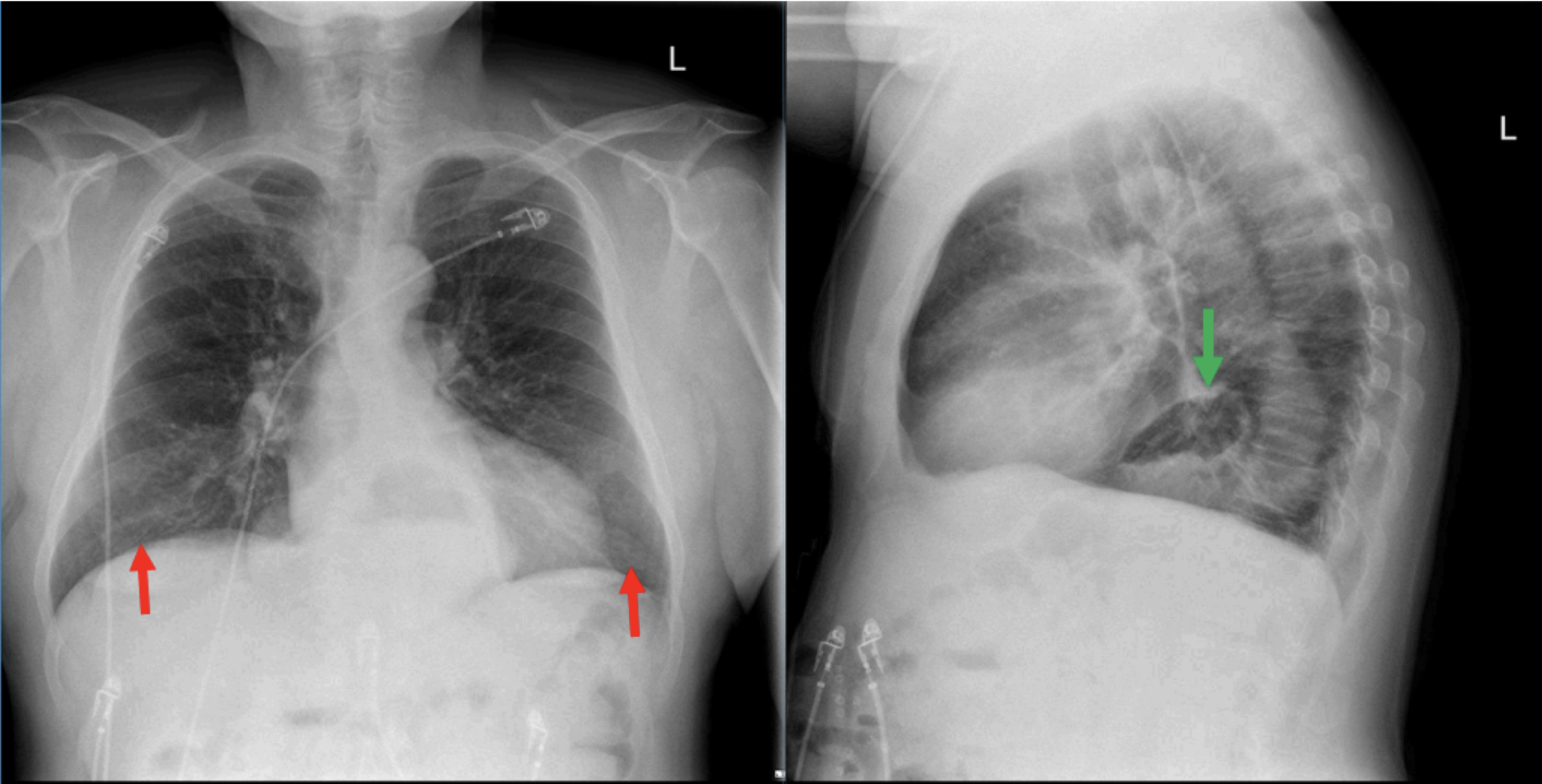
Olive View - UCLA Medical Center. The four major types of hiatal hernia repairs are described with regard to the surgical procedures postoperative radiological manifestations and differential features. Following a diaphragmatic crural repair no subdiaphragmatic esophagus is seen.
Abstract A questionaire regarding the incidence of hiatal hernia in infants and children was sent to the 50 largest departments of pediatric radiology in the United States and Canada. Hiatal Hernia Pediatric A hiatal hernia occurs when a part of the stomach pushes up through the diaphragm. The 36 returned revealed that the larger departments see an average of.
Understanding the expected postsurgical imaging features of these common esophageal surgeries and postoperative complications is essential. Laparoscopic herniorrhaphy and Nissen fundoplication were performed. Two recurrent hiatal hernias were identified on surveillance imaging for a recurrence rate of 43 at a mean 18 10 months after initial operation.

Pdf Hiatal Hernia In Pediatric Patients Laparoscopic Versus Open Approaches Semantic Scholar
Indian Pediatrics Editorial
Rnpmxyfo1lsefm
Hiatal Hernia Pediatric Radiology Reference Article Pediatric Imaging Pedsimaging
Hiatal Hernia Imaging

A Gastrografin Contrast Study Confirming A Paraesophageal Hiatal Download Scientific Diagram
Hiatus Hernia Hong Kong Pdf Ppt Case Reports Symptoms Treatment
Transthoracic Hiatal Hernia Repair Basicmedical Key

Hiatal Hernia Presenting As Difficulty In Inserting Feeding Tube In A Neonate Born Preterm The Journal Of Pediatrics

Malpositioned Nasogastric Tube In Hiatus Hernia Radiology Case Radiopaedia Org

Hiatus Hernia Radiology Reference Article Radiopaedia Org

Congenital Paraesophageal Hiatal Hernia Pitfalls In The Diagnosis And Treatment Journal Of Pediatric Surgery
Cureus Gastric Volvulus A Complication Of Hiatal Hernia

Sliding Hiatus Hernia Radiology Case Radiopaedia Org

Pdf Congenital Hiatus Hernia A Case Series Semantic Scholar

Hiatal Hernia Pediatric Radiology Reference Article Pediatric Imaging Pedsimaging

Radiology Case Hiatal Hernia

Pediatric Hiatal Hernia Radiology Case Radiopaedia Org
Hiatal Hernia Ap And Lateral Chest Radiograph Annotated Jetem 2018 Jetem